Elizabeth Ramsey's Placental Circulation Diagram

Object 5 | This diagram of placental circulation captures a life's worth of research by Dr. Elizabeth Ramsey, who used X-rays and cineradiography to show that maternal blood enters the placenta in "fountain-like spurts," circulates around the fetal blood vessels, and drains back out. Notably, it was illustrated by Ranice W. Crosby, head of the Johns Hopkins Department of Art as Applied to Medicine and the first woman to direct a department at the Johns Hopkins University School of Medicine. #Carnegie125
Ramsey Placenta Drawing by Crosby
Placenta Drawing
The diagram maps how blood flows between mother and baby in five parts: the villous tree, the branching structure through which fetal blood circulates; fetal circulation in cross section; maternal circulation; the pathways of maternal blood through the intervillous space, the region where nutrients and oxygen pass between mother and baby; and the placental margin, where the placenta meets the uterine wall. Ramsey spent nearly 60 years at Carnegie Science and became one of the world's leading authorities on the human placenta. Credit: Carnegie Science / Katy Cain
Back of Ramsey Placenta Drawing
Inscribed on the back of the diagram, Ramsey's own description brings the illustration to life: "Maternal blood enters the intervillous space in fountain-like spurts driven by maternal pressure, this vis a tergo drives the blood in discrete atreams until the momentum is spent. Lateral spread then occurs. Eventually the blood is pushed toward orifices of exit in the placental floor and is drained into maternal veins. 

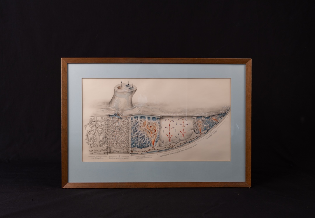
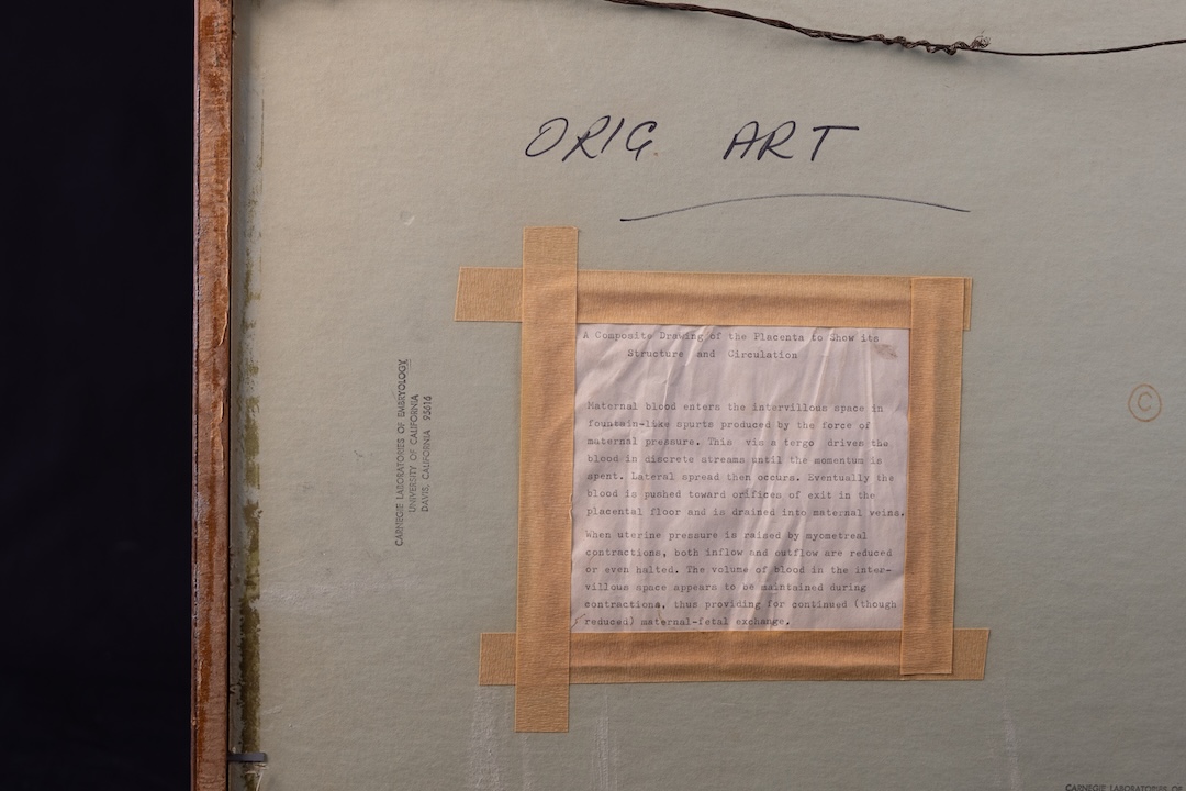
When uterine pressure is raised by myometreal contractions, both inflow and outflow are reduced or even halted. The volume of blood in the intervillous space appear to be mintained during contractions, thus providing for ontinued (though reduced) maternal-fetal exchange." 
Credit: Carnegie Science / Katy Cain