Baltimore, MD— As all school-children learn, cells divide using a process called mitosis, which consists of a number of phases during which duplicate copies of the cell's DNA-containing chromosomes are pulled apart and separated into two distinct cells. Losing or gaining chromosomes during this process can lead to cancer and other diseases, so understanding mitosis is important for developing therapeutic strategies.
New research from a team led by Carnegie's Yixian Zheng focused on one important part of this process. Her results improve our understanding of how cell division gives rise to two daughter cells with an equal complement of chromosomes. It is published by Developmental Cell.
Cell division is helped along by a complex of more than 90 proteins, called a kinetochore, interacting with scaffolding-like structural fibers called microtubules. Together the kinetochore and microtubules provide the structure and force that pull the two duplicate halves of the chromosome apart and direct them to each daughter cell.
By looking beyond the microtubules and kinetochores themselves, Zheng's team identified a protein that regulates the interactions between the kinetochore and the microtubule fibers. Using super resolution microscopy, they were able to hone in on one particular phase of this process, namely the way that microtubules are "captured" by the kinetochore to promote proper alignment of the chromosomes in a way that facilitates equal partition of duplicated DNA.
"The study of mitosis has focused on microtubules and kinetochores, the most prominent structure that researchers observe. Our work demonstrates the importance of expanding the scope of study to include other cellular components because this is critical to achieving an in depth understanding of the mechanisms underlying chromosome alignment in preparation for dividing the DNA into two new cells." Zheng said.
The team included Carnegie's Hao Jiang, the lead author, Shusheng Wang, Junling Jia, and Yihan Wan. Hao Jiang and Yihan Wan are both collaborative postdoctoral fellows in the laboratories of Carnegie's Yixian Zheng and Xueliang Zhu of the Institute of Biochemistry and Cell Biology in Shanghai China.
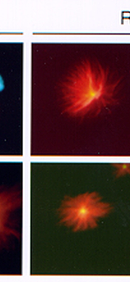
Image Caption: The images show the microtubule-based spindle fibers (red), chromosomes (blue), and their kinetochores (green). Microtubules align chromosomes in the middle of the spindle in the presence of the newly discovered protein called BuGZ (+BuGZ), but mis-aligned in its absence (-BuGZ). Chromosome misalignment leads to its mis-segregation during cell division. Scale bar, 5 microns.
-----------------------
This work was supported by the Chinese Academy of Sciences (XDA01010107), the Ministry of Science and Technology of China (2014CB964803) (X.Z.), the National Science Foundation of China (31010103910) (X.Z. & Y.Z.), and R01 GM056312 (Y.Z.) and R01 GM06023 (Y.Z.).
