San Diego, CA — Ghosts are not your typical cell biology research subjects. But scientists at the Carnegie Institution for Science and the National Institute of Child Health and Human Development (NICHD) who developed a technique to observe muscle stem/progenitor cells migrating within injury sites in live mice, report that “ghost fibers,” remnants of the old extracellular matrix left by dying muscle fibers, guide the cells into position for healing to begin.
Using intravital two-photon imaging combined with second-harmonic generation (SHG) microscopy, the Carnegie’s Micah Webster and Chen-Ming Fan and the NICHD’s Uri Manor and Jennifer Lippincott-Schwartz observed these cells riding to the rescue, using the long axis of these ghost fibers to spread out and orient themselves. The study will be posted online by the journal Cell Stem Cell on Thursday, December 10 ahead of publication in the February 4 issue. Webster will present the ghost fiber work at ASCB 2015 in San Diego on Sunday, December 13.

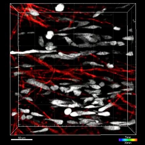
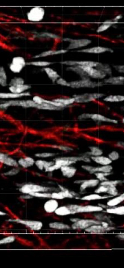
Image: Activated muscle stem/progenitor cells (white) moving within basal lamina remnants, “ghost fibers” (red) which remain when muscle cells degenerate from injury. Courtesy of Micah Webster and team.
This is the first direct visualization of skeletal muscle stem/progenitor cell-mediated regeneration in live mice, says Webster. It also shows the possibilities of combining high-resolution microscopy techniques such as two-photon imaging, which can visualize fluorescent markers at great depth in living tissue with another advanced technique, SHG imaging. SHG turns two photons into one but at half the original wavelength, avoiding problems with dye bleaching and signal saturation.
Combining the two techniques, the researchers were able to image activated muscle stem/progenitor cells moving bi-directionally along the long axis of individual ghost fibers left behind by the lost muscle cells. The stem/progenitor cells spread along the ghost fibers where they could divide, fuse, and fully differentiate into new muscles. When the researchers reoriented the ghost fibers, the regenerated muscle tissue was disorganized. Their direct observation of stem/progenitor cells in action showed ghost fibers playing an “architectural role” in regenerating muscle, says Webster, serving as templates proportioned for laying down new muscle tissue so as to match the same size and align to the same direction as the damaged portion.
