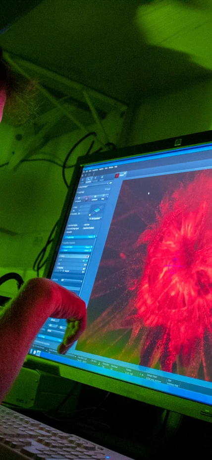
Baltimore, MD — Deep in the Department of Embryology’s dark microscopy suite, Dr. Phillip Cleves studies a magnified image of a sea anemone called Aiptasia, his lab’s primary model organism.
Cleves, who joined Embryology as a Principal Investigator last fall, is trying to help stop the global decline of coral reefs. To do this, he needs to understand what happens—at the cellular and molecular levels—when reef-building corals experience heat stress.
This research is of particular importance because coral reefs are dying at an alarming rate due to climate change. The rising temperature of the world’s oceans is forcing corals to oust the nutrient-supplying photosynthetic algae that live within their cells, a process called “bleaching,” causing the corals to starve. As major biodiversity hotspots, their loss looms large, causing significant damage to global economies and human health.
The symbiotic relationship between corals and their algal tenants is critical for the health of coral reefs—and Cleves believes that understanding the mechanics of this symbiosis could help reefs survive the threat of global warming.

As a postdoctoral researcher at Stanford, Cleves was the first person to develop and apply CRISPR/Cas9 genome-editing technology for use in coral, deploying this cutting-edge tool to study gene function in Acropora millepora from the Great Barrier Reef. His experiments uncovered molecular details of coral biology that had long been poorly understood, due primarily to a historical lack of genetic tools and no tractable model system for coral.
One thing we're really excited about is seeing the bleaching process in real time. We're using Carnegie's fluorescent microscopy to capture what happens at the cellular level as an anemone experiences heat stress.

Aiptasia is a powerful model organism for studying coral biology. Like corals, anemones share a symbiotic relationship with intracellular algae. But unlike corals, they can be easily grown and analyzed in a laboratory.
The image below features two groups of anemones: On the left, healthy Aiptasia with symbiotic algae; on the right, bleached Aiptasia with no symbionts. The damaging effects of heat stress are seen here as a loss of pigment and stunted growth.

With these bleaching experiments, we can recreate what's happening to corals worldwide due to climate change in a small dish of anemones in the lab,” explains Cleves.
Another benefit of using anemones as a model system is that, unlike corals, they can survive without symbionts—as long as they are sufficiently fed. This critical distinction gives researchers the ability to compare the genetic differences between healthy Aiptasia—with and without symbionts—to bleached Aiptasia, healthy coral, and bleached coral, letting them pinpoint exactly which genes and molecular pathways are involved in bleaching.

We can recreate what's happening to corals worldwide due to climate change in a small dish of anemones in the lab.

DID YOU KNOW?
Aiptasia have a superpower: They can regrow parts of their body that have been damaged or lost. This biological phenomenon, called regeneration, allows the separated piece to develop into a whole new anemone, which is a clone of the original! This is called asexual reproduction.

In addition to studying how heat stress affects the genetics of coral-algal symbiosis, the Cleves lab is trying to understand how symbiosis impacts Aiptasia’s mysterious ability to regenerate. This is a new frontier for the lab, so check back soon for details about their research!


Because anemones are soft-bodied animals, they are amenable to some highly sophisticated molecular techniques that require homogenized tissue samples. In the images above, graduate student Amanda Tinoco homogenizes, or grinds up, an Aiptasia to prepare it for gene sequencing. Tinoco, who works in the Cleves Lab through a joint program with Carnegie, Macquarie University, and the Australian Institute for Marine Science, will be extracting RNA from the goo, searching for the genes underlying differences in symbiosis.

Looking to the future, Phillip Cleves stands in what will become his lab’s main grow room. It will house a variety of corals, anemones, and other cnidarian models that share a symbiotic relationship with intracellular algae. By studying each organism's unique biology, Cleves aims to learn more about the unifying principles of symbiosis at large.
To learn more, visit The Cleves Lab
